
Bees Dream of Gold


I chose to study Chemistry and Physics simply because they were the subjects I enjoyed most, so I enrolled on a B.Sc. (Hons) degree at the University of Malta without having a clear idea about what I would be doing once the four years are over. I was not the best brain in the class but in 2004 I graduated with a 2:1 grade and it was quite obvious that I needed a plan. A couple of opportunities to embark on a Ph.D. in Britain came along through local contacts and applications on jobs websites. Despite not knowing much about the subject, I decided to go with the Ph.D. at Exeter University because it was about Nuclear Magnetic Resonance, a subject that sits right on the verge of Chemistry and Physics.
Obviously the idea of moving abroad, living away from my parents and starting this amazing new adventure was incredibly exciting. From the start of my Ph.D. things went incredibly well, it was immediately obvious that I was much better at doing research than studying for exams. I started with looking into dynamics in solid materials on the microsecond timescale, which is the less studied type of motion. It bridges the gap between very fast (spin-lattice relaxation motions, nanosecond) and slow (millisecond to second) timescales. I published my first scientific paper a year into my Ph.D., and five more followed by the time I defended my thesis.
Because of the contacts I built during my Ph.D. as soon as I finished I was offered a post at University College London, Institute of Child Health, working as a research fellow in renal imaging. I carry out research at Great Ormond Street Children’s Hospital using novel non-invasive Magnetic Resonance Imaging (MRI) techniques. I work mainly with children requiring a kidney transplant. The aim of my work is to eventually be able to furnish doctors with information about their patients, which is currently either unavailable to them or they can only get through invasive clinical techniques such as biopsies. My work here has produced six peer-reviewed papers and I am currently working on a few more.
The research I carried out during my Ph.D. involved dealing with basic scientific concepts like Quantum Mechanics — that studies sub-atomic phenomena — and I was at liberty to experiment as I saw fit, which I enjoyed. However, despite being much more restrictive, I find clinical research extremely rewarding. Coming face to face with the people benefiting from all your hard work is really priceless.
Just after my Ph.D. I married my husband. We are now very proud parents of a two-year-old son. Any working mum would tell you that raising a family while maintaining a career is not easy, but I believe that if you like your job enough, combing the two is very worthwhile. Obviously research does not wait for anyone, and luckily for me, having colleagues that supported me meant that I was able to carry on publishing while I was on maternity leave.


 Research — that would be the simplest way to answer the question above. Really and truly this answer would only apply to a small niche of individuals throughout the world.
It is a big challenge to explain to people what you do with a science university degree. The questions “Int għal tabib?” (Are you aiming to become a doctor?) or “Issa x’issir, spiżjar?” (Will you become a pharmacist?) are usually the responses. The thing is, people have trouble understanding non-vocational careers — if you are not becoming a lawyer, an accountant, a doctor or a priest, the concept of your job prospects is quite difficult to grasp for the average Joe.
In truth, it is not really 100% Joe Public’s fault — research is a tough concept to come to terms with, ask a good portion of Ph.D. students about that. There seems to be a lack of clarity in people’s minds about what goes on behind the scenes. If you boil it down, everything we use in our daily lives from mobile phones to hand warmers are the spoils of research — a laborious process with the ultimate goal of increasing our knowledge and, consequently, the utility of our surroundings.
“People need to stop feeling threatened by big words and abstract concepts they cannot grasp”
So, then, why exactly is it such an alien concept? I think the reason is that research is very slow and sometimes very abstract. Gone are the days when a simple experiment meant a novel, ground-breaking discovery — research nowadays delves into highly advanced topics, building on past knowledge to add a little bit more. I have complained about this to many of my colleagues on several occasions — and it is more complicated when you are studying something like Chemistry and Physics, or worse, Maths and Statistics — people just do not get it!
Research is exciting. The challenge is how to infect others with this enthusiasm without coming off as someone without a hint of a social life (just ask my girlfriend). It is nice to see initiatives like the RIDT and Think magazine trying hard to get the message out there that research is a continuous process with often few short-term gains. It can be surprising when you realise how much is really going on at our University, despite its size and budget.
To befriend the general public researchers still need to do more. The first step is relaying the message in the simplest terms possible — people need to stop feeling threatened by big words and abstract concepts they cannot grasp. There also needs to be increased opportunities for interaction with research — Science in the City is the perfect example. Finally, I think MCST needs to start playing a larger role — it must work closer to University and take a more coordinated role at a national level. Only then can we begin to explain what us researchers do.

Producing Food products, pharmaceuticals, and fine chemicals leads to hazardous waste and poses environmental and health risks. For over 20 years, green chemists have been attempting to transform the chemical industries by designing inherently safer and cleaner processes. Continue reading

Classical, romantic, baroque, rock, hip hop… music continues to change throughout the years, yet we all look for that beat that gets us moving. How can we not when music is such an important part of our life?
Music is found everywhere: on television adverts, films, on the radio and at places of worship. Our society immerses us in it for hours every day. A person will listen to music that represents the way they feel. Music has the potential to influence moods, feelings, and thoughts.
“Music opens infinite thinking modes unknown to us and uncovers situations we wouldn’t otherwise experience”
Legendary rock guitarist Jimi Hendrix, told Life magazine in 1969, ‘I can explain everything better through music. You hypnotise people to where they go right back to their natural state, and when you get people at their weakest point, you can preach into their subconscious what we want to say.’
Music, like language, has a common factor: a person’s active role. People create music. No music can exist without the people who make it.
The Ethnomusicologist Dr Philip Ciantar (University of Malta) is interested in both the music itself, as a humanly organised sound, and the musicians. His research focuses on understanding how people worldwide think about music and how that affects their music. He meets and interviews countless musicians and their audiences. People’s thinking about music is shaped by who they are, their world-view, and how they use their creative imagination to create music. Take John Lennon’s song Imagine. The song has touched countless around the world. It might have changed the way people see themselves, relate to the people around them, and influenced future songs.
Ciantar explains that ‘by listening to and exploring music from different countries we can understand other cultural and social realities. Music opens infinite thinking modes unknown to us and uncovers situations we wouldn’t otherwise experience.’ According to him, ‘music can highlight social issues or it can make a connection with different cultures when many other avenues fail’. This is the acceptance of ‘otherness’, the concept of what makes us different from each other culturally and socially. Music can be a very effective medium.
Acceptance of different cultures needs to be taught from a young age. Music can help in showing people the advantages of multiculturalism. Ciantar suggests that, at school, children can be taught instruments used in different cultures. This would help students understand and appreciate not just the instruments but also the musicians playing them. He continues, ‘you need to be open to other opinions, cultures, and traditions’ and music provides the right scenario.
Understanding music globally should lead to appreciation of diverse sounds and how they are made, communicated, and transformed into meaning. The musical process reveals humanity and here otherness surfaces as a challenge for us to deal with. It is up to us to then connect with different cultures we might consider alien.
People come together through music. The village feast is Malta’s best example of unity through music. During a feast a quiet pjazza transforms into a music concert, a fireworks festival, and a food extravaganza — uniting the whole community. These celebrations bring people together ignoring their differences.
Multiculturalism is a worldwide phenomenon. Malta is becoming multicultural and, as Ciantar comments, ‘music is an indicator of what is going on. Performances of African music at the Marsa Open Centre can be interpreted as a plea for social acceptance and cultural integration. Slavic street players in Republic Street play Bach’s violin partitas to make us connect with them culturally. Once we are connected they play a nostalgic lullaby from their homeland to make us feel the pain of distance and sympathise with them. Undoubtedly, music serves as a social text; in itself, an intriguing sonic document that links the evident with the untold or even ignored.’ This is the power of music and the concept of otherness that can shape our thoughts on multiculturalism and readiness to accept others’ views.
“Undoubtedly, music serves as a social text; in itself, an intriguing sonic document that links the evident with the untold or even ignored”

He became even more aware of multiculturalism while conducting his Ph.D. research. He went to Libya to experience different cultural backgrounds and traditions. He worked with Libyan musicians, attending their rehearsals, talked to people on Tripoli’s streets about the musical tradition of ma’lūf (a tradition valued for its Andalusian legacy), and sneaked in percussion performances with Libyan musicians. Apart from writing a book, these experiences helped Ciantar understand otherness and the challenges it implies.
Ciantar’s first experience with ethnomusicology and otherness goes back to 1991, when he was inspired by the writings of John Blacking and Bruno Nettl, and started researching Maltese folk music għana. He saw how the għanejja performed in two different contexts and their music changed accordingly. The music they sang was more elaborate in their regular bars when compared to stage music with an unknown audience.
Otherness can also be scrutinised through musical translation. Ciantar researches musical translation: how we digest and eventually accept music that might not be initially appealing to us. Recently, he composed a Maltese festa band march out of tunes that he had recorded in Libya. The process allowed him to investigate the music and himself. He had to take elements of one musical tradition and apply it to another that was culturally remote, using himself to understand the process of how a person thinks and transforms thought into music.
Ciantar is very hopeful of the musical evolution in Malta as this is being influenced by the different cultures that people encounter everyday. This will create a more varied musical scene. Ciantar can already feel the difference.
Stephanie Mifsud is part of the Department of English Master of Arts programme.
[ct_divider]

Wi-fi is ubiquitous. The technology can be an easy back door for hackers to access a computer through online tools that anyone can learn to use. The global cybercrime bill now tops €700bn and will keep rising. To find out the security of Maltese Wi-Fi networks Kurt Mahoney (supervised by Prof. Ing. Victor Buttigieg) mapped out around 70% of the island’s built-up areas.
Mahoney first carried out in-house testing on Wi-Fi security protocols. He then formulated security categories depending on ease and speed of access to a private network. For example, the WEP (Wired Equivalent Privacy) security standard could be cracked in less than one minute (irrelevant of password complexity). On the other hand, the WPA2 (Wi-Fi Protected Access II) security standard with AES (Advanced Encryption Standard) grade encryption and a twelve-character random alphanumeric password was virtually impossible to crack using brute force techniques.
Setting up a car with several Wi-Fi antennas, he then travelled a preplanned route through all the Maltese villages, apart from Mdina. Private security protocols were noted from automatic Wi-Fi transmissions, however he avoided conducting cracking or penetration testing. Mahoney then created a security map for the Maltese Islands from 64,317 observed private networks. Forty percent of private Wi-Fi networks in Malta were very vulnerable to hacking that increased to 90% if using more sophisticated attacks.
Wi-Fi security was poor all over the Island, with Western and South Eastern districts having marginally lower security. Malta needs a nationwide awareness campaign to increase the security levels of Wi-Fi networks. Top-notch security can be setup in a few minutes. All modern routers support military grade AES encryption standards, and together with a proper password policy one would render a Wi-Fi network almost invulnerable to direct attack.
This research was presented at the fourth Workshop in Information and Communication Technology (WICT). It was performed as part of a B.Sc. (Hons) in ICT at the Faculty of ICT and part sponsored by the Malta Communications Authority. It was shortlisted for the Chamber of Engineers’ Best ICT Student’s Project Award.


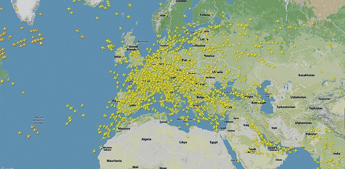
Every day around 30,000 aircraft take to Europe’s skies. Choreographing this airborne dance is daunting. At the moment, it is orchestrated by the disparate air traffic management systems of each European country, with control handed over at border crossings. The aeronautics research team at the University of Malta is part of an ambitious EU project to change that by establishing a single European sky, enabling EU air traffic controllers to manage increasing amounts of traffic with greater safety, lower costs, and a reduced environmental impact.
Ask Prof. Ing. David Zammit-Mangion (Department of Electronic Systems Engineering, UoM) what he loves and he will reply, ‘anything that flies’. He has come a long way since his childhood dreams of flight, when he would build model aeroplanes and scamper over fences to photograph real ones. Now he leads a major research team with an important role in Clean Sky, the EU’s €1.6 billion flagship project which aims to reduce the environmental impact of air transport.
The enthusiasm for flying never left Zammit-Mangion. As an adult, he eventually took to the skies himself, learning to fly during his doctoral research at Cranfield University in the UK, where he designed a cockpit instrument to monitor the take-off performance of aircraft. ‘My dream was to twin my passion with my profession,’ he said. It is a formula that has worked. Zammit-Mangion’s familiarity with commercial operations, safety procedures, and aircraft equipment has given his research an edge by enabling him to quickly estimate the cost and feasibility of different approaches. ‘When it comes to addressing problems, you need to have a very broad understanding of the whole industry,’ he says, and his hands-on industrial experience and hours logged in the cockpit have proven invaluable. Clean Sky is central to meeting the environmental goals embedded in the vision of a unified European sky. Launched in 2008, its goal is to reduce the excess noise and greenhouse gas emissions created by aeroplanes. Air transport is responsible for around 2% of global carbon dioxide (CO2) emissions, but traffic is expected to more than double by 2030. By improving air traffic management (ATM) and aircraft technology, the 600-member Clean Sky project aims to ensure that emissions increase at a slower rate than demand.
Aeroplanes currently follow flight paths through set air corridors, which can make routes unnecessarily long. They also may have to climb or descend in stages and wait in a holding pattern at their destination. These inefficient practices increase the amount of fuel used, leading to higher costs and greater greenhouse gas emissions. Each kilogram of jet fuel burned releases roughly three kilograms of CO2 into the atmosphere, along with other greenhouse gases like nitrogen oxides. This happens high in the atmosphere, where these gases end up taking part in a variety of physical and chemical processes that cause them to have a greater environmental impact than they would closer to the ground. Given that many airliners burn around 50 kg of fuel per minute, even relatively small optimisations can have a significant impact.
“Each kilogram of jet fuel burned releases three kilograms of CO2 into the atmosphere, along with other greenhouse gases like nitrogen oxides”
Improving air travel routes is not a simple task. It is what engineers call a ‘multi-criterion, multi-parameter problem’. In other words, you have to balance lots of factors, like the type and mass of the aeroplane, weather conditions, route limitations, and air traffic control constraints. At the same time, you need to maximise performance on different objectives such as fuel use, flight time, and environmental impact. Zammit-Mangion describes it as ‘a very complex mathematical problem’. That sort of complexity might sound like a nightmare to most people, but it is just the sort of thing Ing. Kenneth Chircop thrives on. ‘My real love is for engineering mathematics,’ said Chircop. He studied engineering for his degree, but then his passion for mathematical challenges drove him to join the aeronautics research team. ‘At the end of the day, I wanted to do something heavy in mathematics again.’ As their contribution to Clean Sky, the team developed a software package called Green Aircraft Trajectories under ATM Constraints (GATAC) to help optimise flight routes. Instead of just performing a single optimisation, GATAC provides an optimisation framework which aircraft operators can use with their own models. By plugging in models of aircraft and engine performance, emissions levels, noise production, and so on, users can work out optimal air travel trajectories to match their constraints and conditions. The core software developed at UoM incorporates various models from different research partners, but users are also free to plug in their own models. Aircraft manufacturer Airbus uses GATAC with its own proprietary models. ‘It’s great to see that foreign partners look at us as equals,’ said Chircop. ‘They trust us to develop state-of-the-art technology. We have delivered, and they trust us to keep delivering. We’re really proud of that; it’s what makes us tick and want to do more.’

This work has brought more than just international recognition to Malta; the country will also enjoy practical benefits. Kenneth Chircop is spear-heading Clean Flight — a national research project financed by the Malta Council for Science and Technology’s national research and innovation programme 2011 — to apply the lessons from Clean Sky to Maltese airspace. ‘Our impact on the national scene can be remarkable,’ said Chircop, describing the gains to be made by optimising the arrival and departure routes aeroplanes use at Malta airport. As an island nation, Malta relies heavily on air traffic to connect it to the rest of the world. In 2013, Malta International Airport saw over 30,000 arrivals and departures, up from roughly 26,000 only seven years ago. Despite this, its air traffic systems need an overhaul; while the technology is state-of-the-art, some of the procedures are out of date. For example, aeroplanes arriving and departing from an airport follow standard, published routes, called STARs (Standard Terminal Arrival Route) and SIDs (Standard Instrument Departures) respectively, which can simplify airspace management. ‘The SIDs in Maltese airspace were designed years ago when fuel was relatively cheap, and the impact combustion made on the environment was not given due importance,’ said Chircop, ‘and we don’t even have STARs.’ Updating these procedures presented a clear opportunity to reduce fuel use and greenhouse gas emissions in Maltese airspace. Together with their partner, Maltese aeronautics consultancy company QuAero Ltd, Chircop, Zammit-Mangion, and the rest of the team analysed the flight paths taken by aircraft in Maltese airspace and discovered that they were scattered and inefficient. They developed a tool to design and analyse the best arrival and departure routes for aeroplanes, which they used to calculate revised routes for Malta’s airport. Based on fuel savings estimates for the Boeing 737 and Airbus A320, the two most common aircraft in Maltese airspace, the new routes could save 465 tonnes of fuel for departing aircraft and 200 tonnes for arrivals every year. The fuel reductions mean less money spent and lower CO2 emissions in Maltese airspace. Not only does that directly benefit Malta’s environment, but it also offers indirect benefits by reducing the pressure on Malta’s carbon emission caps. In addition to improving the course followed by flights, the team has helped improve climbs and descents. Planes can approach the airport in many different ways: for example, a smooth, continuous descent, a series of steps interrupted by level flight, or a close approach at full altitude followed by a quick descent. Determining which approach is optimal is a dynamic problem that has to factor in the weight of aeroplane and its cargo, weather conditions, operational constraints, air traffic and so forth. Current optimisation methods try to balance flight time and fuel use, but do not take the other factors into account. The Clean Flight team developed a new approach using computer algorithms which can improve the efficiency of climbs and descents in around 10 minutes on a single computer. ‘So 15 minutes before departure, for example, an air traffic controller can calculate the optimal route for the flight at the current conditions,’ said Chircop. Altogether, this work could save 1,500 tonnes of fuel every year.
The sky is the limit for this aeronautics team. As Clean Sky winds to a close, the EU is preparing to launch Clean Sky 2, and the UoM team will probably continue to play a significant role in the initiative. On the national front, the optimisation system developed in the Clean Flight project will be tested with actual flight trials over the coming months – a major step forward in a field where such tests are incredibly expensive and safety is always a paramount concern. According to Chircop, it is an indication that the potential benefits are large. ‘We’re pushing to get this technology into the field so we can see it making actual gains, instead of simply on paper,” he said. Meanwhile, the GATAC software package is already being used by key industrial players, according to Zammit-Mangion. Looking forward, it clearly has a scope beyond Clean Sky, and may even come to be used by other industries like maritime shipping, which faces similar problems. The team is also working on a project to test unmanned aerial vehicles (UAV) flying with commercial aircraft in an air traffic control environment. Although the UAV tech was developed in Italy, the Maltese team will test its operational aspects. If successful, the project could open the door to the integration of UAVs into the wider aviation community. The aeronautics team has put Malta on the map when it comes to aviation research, a major accomplishment for a nation with no significant track record in the field until ten years ago. ‘We’re well-established and recognised in European and global research circles,’ said Zammit-Mangion, describing the team’s success. With the network of partners they have built up and the quality of the team’s research, the future is looking up.
[ct_divider]
Dr Sedeer El-Showk is a freelance science writer. He blogs at Inspiring Science and for Nature’s Scitable network.


Why should public, private, and non-profit entities invest in research? Several reasons exist.
Firstly, research is key for our future. Research helps drive new knowledge that will improve the world. Society depends on research, across a wide range of disciplines, to strengthen our quality of life and sustain economic stability. By raising funds for University research the RIDT is surely not trying to reinvent the wheel. On the contrary, throughout Europe, the US and Asia public universities are enhancing their Government funding through various initiatives to sustain important research. Universities all across the globe appeal to public entities, private individuals and NGOs to fund research and invest in our society’s future.
Locally, we have just started scraping the surface of fundraising for research. Recently, the RIDT received a number of important donations which shall serve to continue fostering local research. In the first of its kind, we received €55,000 from local NGO ‘Action for Breast Cancer Foundation’ (ABCF), raised through the ALIVE Cycling Challenge. They are being used to launch a Ph.D. studentship in breast cancer research. The Lifecycle Foundation has also donated €70,000 towards kidney disease research. These NGOs have followed a stream of public and private entities, as well as students, who have been donating money for research for the last three years.
Secondly, don’t you want to be part of something bigger? You probably cannot find the cure for cancer yourself, but everyone can contribute to make that a possibility. As the University’s Research Trust, we do not only want to attract big corporate companies or NGOs to donate money, but we also want you — the students, the alumni, the professionals, the workers, the parents, to realise that donating for research is a noble cause. A recent Christmas campaign at the University of Malta that was spearheaded by KSU, the UoM staff and the Chaplaincy managed to raise €12,000 from students, staff, and academics on campus. A third of these funds were donated to the RIDT, which were devolved to the Department of Anatomy. They will be invested in specialised research projects focusing on specific strands of cancer, such as leukaemias, sarcomas, brain tumours, breast and colon cancer.
Research affects our day-to-day lives. Though research discoveries take time and need constant investment to benefit our society, we can come together as a Maltese community by investing in research for good causes. Ultimately, let us imagine a world where we have cured all major diseases, where we can move objects with our thoughts, and unravel the mysteries of the universe.Imagine, and let’s make it happen!

Made infamous by Sigmund Freud, the idea is that we spend one third of our lives dreaming about what we would like to do. Our rational brain suppresses these feelings.
On the other extreme, our brain is just as active in certain sleep stages. These neural firings express themselves in dreams. There are no deep hidden emotions behind them.
Somewhere in between lie recent studies that show that dreams are important in memory, learning and emotions. If you sleep without dreaming these qualities will suffer. For example, rat studies in 2001 showed how while dreaming they replayed solutions to mazes to commit them to their long-term memory.