Fruit flies are not human. Yet they are close enough to have been used for over 100 years by scientists to find out more about humans. Dr Ruben J. Cauchi writes about his relationship with the fly. He uses it to find out how to stop Alzheimer’s disease, Parkinson’s disease, and Motor Neuron Disease that affect tens of millions
It was a cold and grey February afternoon. Snowflakes were pelting the dreaming spires of Oxford. This gloomy weather did nothing to impede the warmth and buzz exuding from the laboratories crammed in the iconic Sherrington building. Less than a century earlier, this labyrinthine edifice was the habitat of Sir Charles Sherrington whose experiments shaped our understanding of the ‘synapse’ or the minute gaps between one brain cell (neuron) and another. The Sherrington building (part of the Department of Physiology, Anatomy, and Genetics at Oxford University) has undergone several expansions over the years. In its newest wing, nowadays it houses the research group of Dr Ji-Long Liu, a rising star in the field of genetics and cell biology.
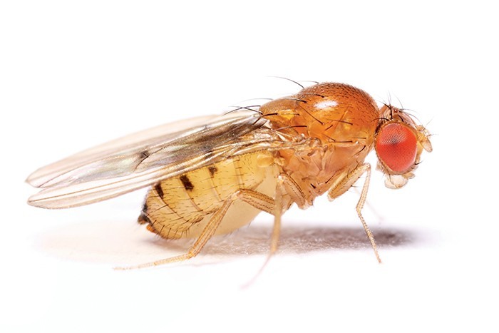
For me, this was no ordinary afternoon. Together with Liu’s lab teammates, I was perched on a stereomicroscope whilst holding a delicate brush in my hands. On one side was a tray jammed with vials populated with fruit flies and the usual good strong cuppa. Fruit flies are no house flies: each adult fly is only a few millimetres long, their beautiful bodies are pale with black zebra-like stripes and their eyes a bright apple-red colour. I grabbed a vial, fired a puff of carbon dioxide gas through its fluffy plug and then firmly rapped the upended vial to shake its sleepy occupants onto an illuminated pad. I took a deep breath before peering at them through the eyepieces.
At the time, I was more than mid-way through my doctoral studies, and the results of my experiments were far from extraordinary. I was researching the most common genetic killer of human infants, a neuromuscular degenerative disease known as spinal muscular atrophy or SMA in short. I was exploiting the tiny fruit fly to gain new insight into this catastrophic disease.
I decided to up my efforts by generating a series of mutants or faults in Gemin3, the gene that I was investigating. I was targeting these mutants to different organs such as brain, muscle, or gut. The results of this screen were due today. With a few flicks, I deftly flipped and sorted the minuscule fly bodies into neat piles taking note of differences that are invisible to the untrained eye. The mutants did not produce any dramatic effect. Damn! Another experiment down the drain! Frustrated by the result, I mistakenly knocked over a vial, dislodging its plug. Usually, released flies would happily escape by flying. Strangely, my flies were jumping as if attempting flight but just couldn’t make it into the air — an unexpected but interesting trait or phenotype. I checked the tag on the vial. In these flies the mutant was targeted to that part of the body that powers movement, the so-called ‘motor unit’. Following that afternoon, which will remain forever etched in my memory, the results just flowed in and a few months down the line I would find myself donning my subfusc (Oxford-speak for academic dress) to defend my doctorate.
Fly Superstar
The rise to biological stardom for the fruit fly, scientifically known as Drosophila melanogaster, began in 1907 when my great-great-grandfather (by academic lineage) Thomas Hunt Morgan adopted this organism to understand heredity or genetics. Morgan was the first to harness the major advantages of working with this organism: they have an insatiable sexual appetite and a speedy development (only 10 days) from embryo to adult. This means that large-scale experiments are doable in record time. Morgan’s infamous ‘Fly Room’ at Columbia University in New York set the stage for a new ‘religion’ practiced and preached across the globe.
Morgan spent years searching unsuccessfully for flies with clear, heritable differences so that he could investigate how they are inherited. A breakthrough happened in April 1910 when he discovered his first mutant, a white-eyed male fly amongst many red-eyed flies. Morgan took great care of this special fly: he kept it in a bottle and after a day’s lab work he used to take it home! At the same time his wife Lilian, who also became a famous geneticist, gave birth to a child. And such was the excitement surrounding Morgan’s discovery that on his first visit to the hospital, Morgan’s wife said: ‘How’s the fly?’ To which, Morgan replied: ‘How’s the baby?’.
When the white-eyed fly was bred or crossed with a virgin red-eyed female, their offspring were all red-eyed. When sisters and brothers were crossed, half of the male progeny gained back their white-eye colour. This hereditary pattern is typical for a sex-linked (recessive) variation, since the gene for eye colour in Drosophila, named by Morgan as the white gene, is on the X chromosome which determines sex. Similar to us, male flies are XY whereas females are XX. This key experiment and numerous others that followed expanded on the knowledge gained through the ingenious cross-breeding experiments of pea plants by the Austrian monk Gregor Mendel half a century earlier. Importantly, this fly-based work found that characteristics like eye colour are inherited from parents through chromosomes — large structures which package DNA in our cells. Furthermore, Morgan and his gifted students uncovered that the thousands of genes in our genome are arranged along chromosomes in a precise order, like beads in a necklace. Each gene can be identified by its specific location on a chromosome.
“Flies could be used as models of human disease”
In 1933, Morgan won the Nobel Prize for these great discoveries. The first of six awards was to recognise seminal insights into our biology through this tiny fly. Hence, in 1946 one of Morgan’s protégés, Hermann Muller, was recognised for his fly research demonstrating that X-rays can damage chromosomes. Then in 1995, Ed Lewis, Christiane Nüsslein-Volhard, and Eric Wieschaus shared the Nobel Prize for their herculean efforts in discovering the genes that controlled early development in Drosophila. In the embryo, waves of master genes are triggered that lead to eyes, brains, and the body’s patterning. Similar genes were later found in humans doing the same function. In 2011 Jules Hoffman received the Nobel Prize for finding how the body’s inbuilt immunity works through the use of the fly model organism. I suspect that there is still room for more trophies in the fly triumph cabinet.
At the dawn of this century, the genomics revolution led to the complete DNA sequencing of an organism including fly and human. These monumental projects revealed that an astonishing number (more than two-thirds) of human genes involved in disease have counterparts in the fly. This development meant that flies could be used as models of human disease. It sparked off a renaissance of Drosophila research. The fly was good at modelling neuro-degenerative conditions because their nervous system has stunning similarities to ours. Neuro-degenerative diseases including Alzheimer’s, Parkinson’s, Huntington’s, and Motor Neuron Disease occur when neurons in the brain and spinal cord begin to die slowly. Patients may lose their ability to function independently or think clearly. Symptoms progressively worsen and ultimately, many die. Most neuro-degenerative diseases strike later in life, so we should expect their frequency to soar as our population ages — Alzheimer’s disease may triple in the US alone by 2050.
Malta: the right time to fly?
Together with my students in my lab at the University of Malta I am working with flies to learn more about neuro-degenerative disease. We continue to focus on SMA, a genetic disorder arising from the deterioration of motor neurons which are nerves that communicate with and control voluntary muscles. As the motor neurons die, the muscles weaken with drastic effect on the walking, crawling, breathing, swallowing, and head and neck control of unfortunate children afflicted by this condition. The child’s intellectual capacity is unaffected but vulnerability to pneumonia and respiratory failure means that many patients die a few years after diagnosis.
The underlying cause of SMA is usually a gene flaw that results in low levels of a protein called SMN for survival of motor neurons. Inside cells, SMN is bound to other proteins called Gemins. The SMN-Gemins alliance is involved in building the spliceosome, which is the chief editor of messenger RNA molecules. Messenger RNA carry the DNA code that instruct cells how to fabricate proteins. If SMN is absent spliceosomes do not form, correctly-edited messenger RNA are not produced and protein synthesis is heavily disrupted — the cell should shut down. Spliceosomes are required in each of the 120 trillion cells forming our body. Yet, in the disease SMA only motor neurons die. The reason has baffled researchers for decades and remains unsolved.
Is it possible that SMN has another function in motor neurons? And does it act alone? Our flies were crucial in providing some answers to these questions. Our work showed how the SMN-Gemins family is tightly-knit. In this regard, we recently demonstrated that both SMN and Gemins can be detected in prominent spherical specks in different cellular compartments. Within the cytoplasm, these organelles are known as U bodies because they probably are the factories of spliceosome components, which themselves are rich in the chemical Uridine. In the nucleus, the structures containing the SMN-Gemins family hug the mysterious Cajal bodies — discovered over a century ago by Spanish Nobel laureate Santiago Ramón y Cajal.
“We are feeding these flies the Mediterranean diet derivatives to see whether Alzheimer’s can be stopped in flies, which will bring us one step closer to treating it in humans”
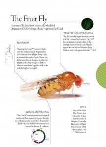
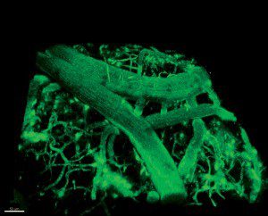
And what about the flightless flies? Think about it. Considering that SMA is a neuromuscular disease, it makes perfect sense that on loss of SMN, muscles become so weak that flies are unable to flap their tiny wings fast enough to fly. Our latest work reveals that flightlessness is seen in flies without enough Gemin proteins. This means that SMN does not function alone but hand in hand with the Gemins. Our next step was to find out the pathway connecting the SMN-Gemins family to the motor defects. We linked the Gemin mutant which did not work properly to a tag called green fluorescent protein or GFP. GFP glows under the right light in cells. We managed to create genetically-modified flies with this modified gene — a first for Malta and a powerful tool to solve the mysteries of this disease.
Fluorescent proteins let researchers figure out a protein’s location. And by knowing the location of proteins we gain of lot of information about what they do. Consider this analogy with a VIP. If we tagged the Prime Minister of Malta we would find that he is most probably found in Valletta most time of the year. If we were aliens from another planet, this knowledge would allow us to refine our understanding of the Prime Minister’s function. Therefore, we can eliminate a function in the entertainment industry (weak signal from Paceville) but we cannot exclude a function in government (strong signal from Valletta). Likewise, we found that our GFP-Gemin mutant is mostly found in the cell’s nucleus. The nucleus houses life’s instruction manual: DNA. Our work now needs to zero in on the other proteins the SMN-Gemins family works with in the nucleus. Doing so will open new therapies to halt neuro-degeneration in children. Back to our analogy, we need to zoom in on Valletta until Auberge de Castille, the Prime Minister’s office, is clearly in focus.
 Several neuro-degenerative diseases occur because of sticky protein clumps that wreak havoc inside, and outside, neurons. This is typical in Alzheimer’s disease, Parkinson’s disease and Motor Neuron Disease. With Dr Neville Vassallo’s research group, and local industry (Institute of Cellular Pharmacology), we are testing chemical derivatives of the Mediterranean diet and flora on fruit flies to see whether they can curb the protein clumps’ toxicity. They definitely do in a test tube. Flies mutated to be remarkably similar to human Alzheimer’s lose their ability to climb up the sides of their vial habitats and die prematurely because of neuro-degeneration. We are feeding these flies the Mediterranean diet derivatives to see whether Alzheimer’s can be stopped in flies, which will bring us one step closer to treating it in humans.
Through flies we have understood human biology. Apart from choosing Mr and Mrs Right, a good geneticist must learn to focus and listen to what flies are really saying. This is easier said than done but achievable. Flies have spurred me to pursue unexpected but interesting paths. In the years to come I, together with my students, will continue to flip, sort, screen and tag, looking for fly mutants who will continue to teach us about ourselves. And yes, we will be all ears!
The author is indebted to colleagues at the UoM and worldwide for their constant support and inspiration. The research of Dr Ruben Cauchi (Department of Physiology & Biochemistry, UoM) is funded by the Faculty of Medicine and Surgery, the University of Malta Research Fund and the Malta Council for Science & Technology (MCST) through the National R&I Programme 2012 (Project R&I-2012-066). For more about Dr Cauchi’s research click here.
[ct_divider]
Find out more:

