

Tumours are not discreet. As cancer cells grow and change, they release microscopic particles into their environment. Some of these particles enter the bloodstream. They are small, abundant, and, as researchers are beginning to realise, potentially informative. A Maltese research project is testing whether extracellular vesicles can provide a less invasive source of cancer information.
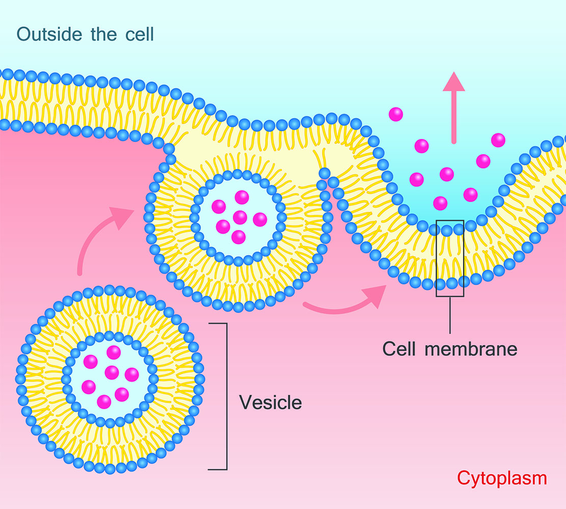
A research group at the University of Malta is investigating how much information these particles can provide. V-MELD, a project funded by Xjenza Malta’s Research Excellence Programme, focuses on extracellular vesicles (EVs): microscopic, membrane-bound parcels released by cells, including cancer cells. These vesicles matter because they can carry molecular characteristics of their cells of origin.
Dr Laura Grech, a resident academic at UM’s Faculty of Health Sciences, describes them as ‘tiny packages released by cells that carry biological information about their cell of origin.’ The question behind V-MELD is whether those packages can be intercepted and read.
For now, the researchers are starting with a simpler system than blood: cancer cells cultured in the laboratory. This is a deliberate choice. Blood is a complex mixture, containing cells, proteins, and particles from across the body, including EVs from healthy tissues. To identify a cancer-specific signal within this background, researchers must first establish what to look for.
V-MELD began by studying lung-cancer cell lines under controlled laboratory conditions. Researchers collected the EVs produced by these cells and analysed the molecular markers present on their surfaces. The initial objective was not diagnosis, but to establish proof of concept: whether researchers could isolate vesicles and reliably detect combinations of surface markers.
The distinction is important. Liquid biopsies, tests that seek information about cancer from blood or other body fluids, have become an attractive area of oncology research partly because ordinary tumour biopsies have an obvious drawback – they require tumour tissue.
Such tissue remains essential. Imaging, biopsies, molecular testing and genetic analysis are already central to cancer care, and V-MELD is not intended to replace them. The potential value of EVs lies in their ability to provide additional evidence without repeated tissue sampling.
‘The idea behind EV research is therefore not necessarily to replace these established techniques, but potentially to complement them,’ says Grech.
Reading the Parcels
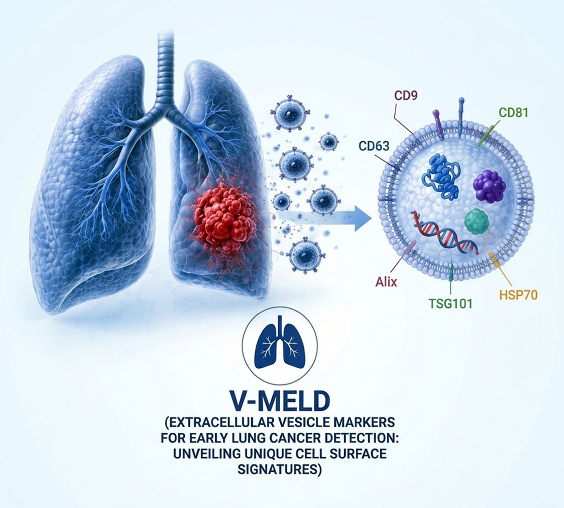
This possibility depends on the vesicle’s molecular composition. EVs display various molecules on their outer membranes. V-MELD treats some of these as identification tags. By targeting specific tags, researchers can isolate groups of vesicles and analyse the additional markers they present.
The relevant information may reside not in a single marker, but in a particular combination.
This is significant because cancer is highly heterogeneous, meaning that two tumours with the same diagnosis may differ biologically. Even among the lung-cancer cell lines studied by V-MELD, vesicles did not always display identical patterns of surface markers. Such variation is not simply experimental noise; it may reflect the underlying heterogeneity that complicates cancer characterisation.
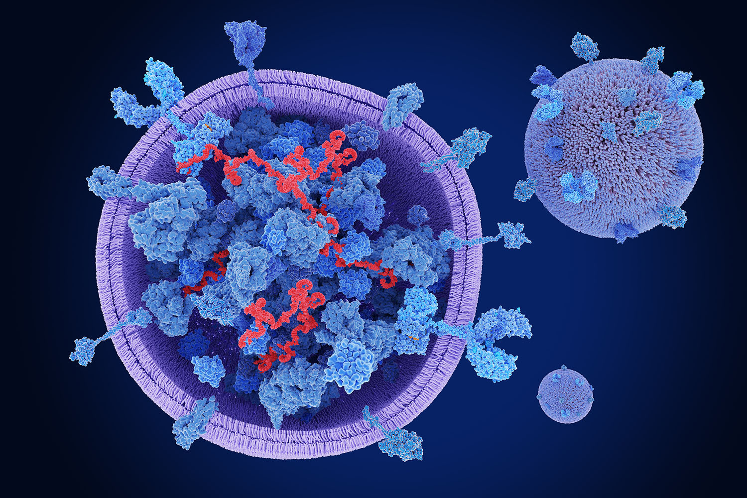
V-MELD is therefore searching for patterns rather than a single molecular giveaway. Grech compares the process to recognising a person by several characteristics rather than relying on only one. ‘The more relevant pieces of information we combine, the clearer the overall picture may become.’
Watching Cancer Change
This approach may prove especially valuable if EVs can eventually be used to monitor changes in cancer over time. A conventional biopsy yields detailed information, but only from a single location at a single time point. Tumours, however, continue to evolve, especially during treatment.
Meanwhile, cancer cells keep releasing vesicles. If researchers can reliably identify tumour-associated EVs in blood, repeated sampling could let them observe changes in cancer without repeated tissue biopsies. This may then make it possible to track shifts in EV patterns as cancer progresses, responds to therapy, or develops resistance.
This remains a future application. V-MELD is still establishing the biological and technical groundwork necessary to address such questions in patients.
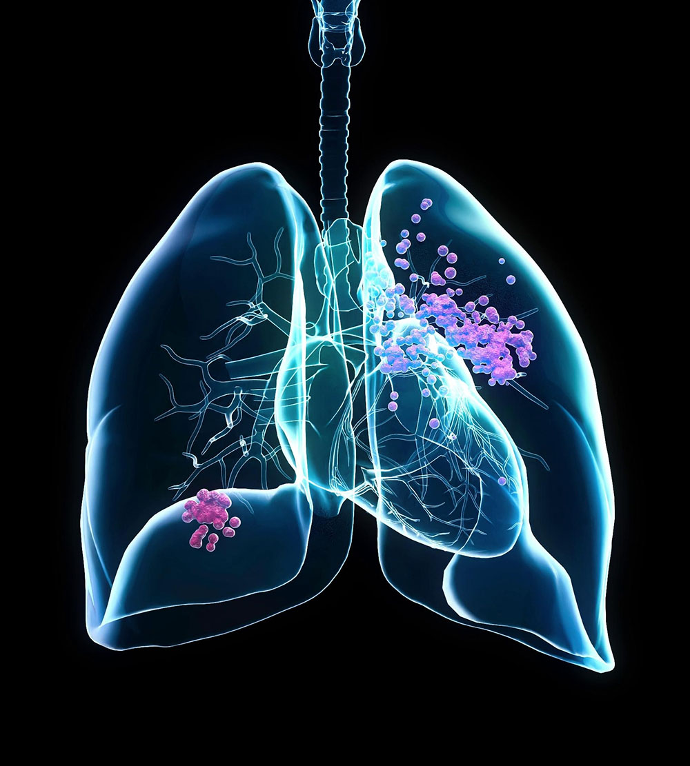
A similar limitation applies to early detection. V-MELD chose lung cancer as its initial model partly because early detection matters. Early disease can cause no obvious symptoms, while complaints such as persistent coughing, tiredness or shortness of breath can have many explanations. Some cancers are therefore diagnosed only when they have progressed further and become harder to treat.
A reliable blood-based pattern associated with cancer would be valuable. However, V-MELD is not currently an early-detection test. Its current aim is to determine whether EVs contain consistent and meaningful signals that could support such applications in the future.
Bridging this gap will be challenging. EVs are both very small and highly diverse. Even under controlled conditions, different cancer cell lines release different EV populations. In blood, researchers must also contend with vesicles from healthy tissues and other biological components.
The challenge is not merely to detect EVs, but to consistently identify the relevant ones.
‘How do we reliably identify the EVs that are actually providing useful information about the cancer?’ asks Grech. Standardisation is central to the work. Sample collection, storage, isolation and measurement can all alter results. V-MELD follows internationally recognised recommendations for EV research to make experiments consistent and findings reproducible by other laboratories.
Clinical diagnostics require that tests yield consistent results, regardless of who performs them or where. Methods that work only in the originating laboratory are of limited value.
From Bench to Blood
V-MELD is not there yet. The next major step is to move from cultured cells to patient samples and ask whether patterns found in the laboratory can also be detected reliably in blood. Promising signals would then need to be tested in much larger groups of patients and in suitable control groups. Researchers would have to show not merely that the technology works, but that it provides information which is accurate, reproducible and clinically useful. Only after extensive validation would regulatory approval for a diagnostic test become relevant.
Nor need the idea stop with lung cancer. EVs are released by many tumour types, and the methods developed through V-MELD could in principle be adapted to others. Pancreatic cancer, another disease in which early diagnosis is particularly difficult, is already one area of interest. The longer-term ambition is broader still: to determine whether the principles established in V-MELD can contribute to a liquid-biopsy platform applicable across several cancers.
Such an application remains far from routine clinical use. The approach’s appeal is clear. As cancer grows, it releases biological material into the body. If researchers can determine which signals are informative, a blood sample could eventually provide insight into a tumour without invasive procedures.
For now, V-MELD focuses on foundational work: developing methods to isolate vesicles, identify informative molecular patterns and understand what they may reveal about the cells that released them.
Project V-MELD (Extracellular vesicle markers for early lung cancer detection: Unveiling unique cell surface signatures) is financed by Xjenza Malta through the Research Excellence Programme (REP-2024-029).




Comments are closed for this article!