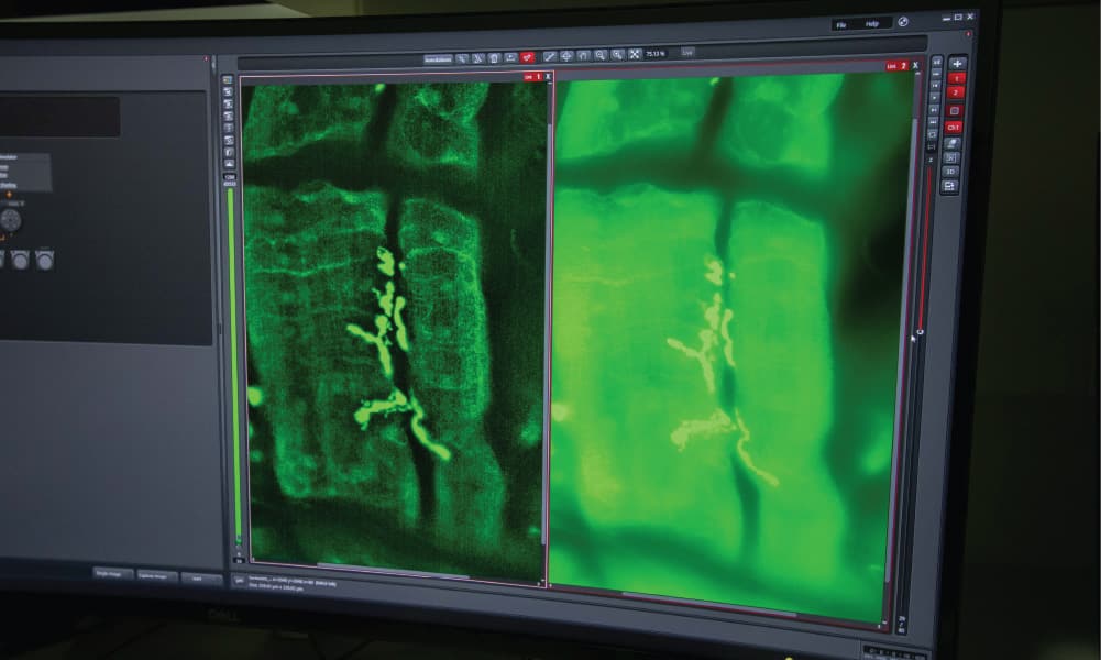
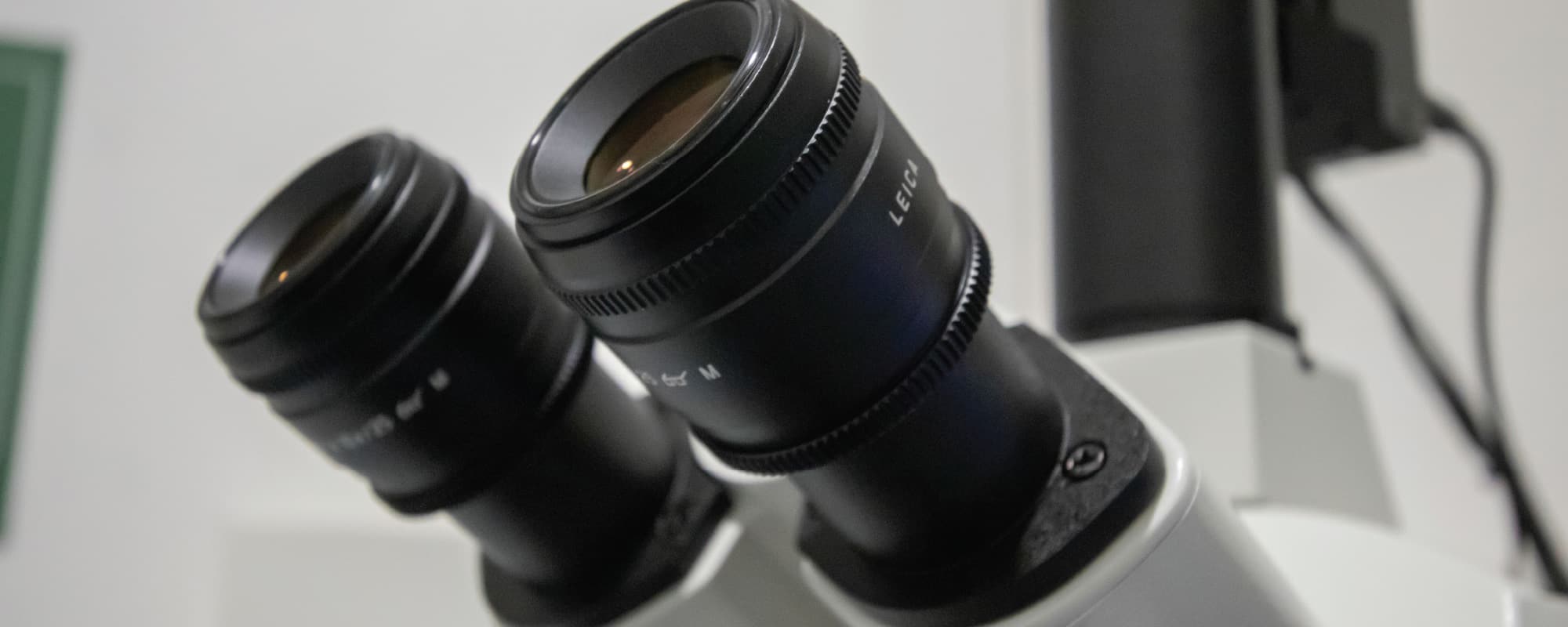
The Leica Thunder Imaging System not only lives up to its grandiose name, it also exceeds it in its purpose. At first glance, the system looks just like your traditional microscope with a flatscreen monitor connected to it. THINK finds itself in the Motor Neuron Disease laboratory at the Centre for Molecular Medicine and Biobanking at the University of Malta. The dim lighting around the setup creates an impressive atmosphere. Images flash on the attached screen, and the sophisticated high-tech features of this specialised microscope become clearer. ‘There is no other microscope like this,’ says Mr Zachary Muscat, Accounts Manager at Evolve Ltd. — suppliers of this equipment to the University.
While traditional microscopes have no issue focusing on normal cells, they tend to struggle with tissue samples. Tissue samples are somewhat thicker, and a typical microscope causes blurring at the centre of the projected image. Clarity and sharpness are critical in a field that requires precise analysis of samples, and the distortions caused by such image processing can severely limit the researcher.
Being only one out of a hundred currently in use worldwide, the Leica Thunder Imaging System is capable of removing this blurring in real-time. Prof. Ruben J Cauchi, who leads the laboratory, explains the concept behind this piece of technology. ‘It looks like a normal microscope,’ he says. ‘The difference is that it has a tower with a powerful processor, and this is its core facility.’ Its high processing power, combined with technology typically used for gaming, allows for the enhancement of images beyond the capabilities of a standard microscope. While traditional microscopes use natural light, the Leica Thunder Imaging System splits natural light into different wavelengths to excite different fluorescent stains, and the processor captures the illumination of these stains independently, while the software compiles the images to produce razor-sharp results.
The laboratory’s primary research focuses on motor neuron diseases such as ALS (Amyotrophic Lateral Sclerosis), by utilising fruit flies as specimens under the microscope’s powerful lens. These insects serve as a model organism of ALS by removing genes causing the disease. ALS flies typically end up with weakness of the muscles used for flight. Prof. Cauchi emphasises the impact of the Thunder microscope for such research. ‘What we can do now is dissect the organism and see what is actually happening at a molecular level in the neurons and muscles. Previously, that was difficult to do.’

Besides ALS, the laboratory is also focusing on projects concerning COVID-19. Research is being conducted on the ACE2 receptor, that same receptor which coronavirus particles bind themselves to before entering human cells. ‘So with the microscope, we are also looking at the location of this receptor and how we can actually find therapeutic approaches that decrease the levels of this receptor.’ In the long run, this will have a significant impact on the health sector by providing it with crucial information for the creation of specific drugs which can be used not only for COVID-19 but also potentially for future pandemics.
Equipment supplied by Evolve Ltd. through collaboration with the University of Malta, and made possible with funding from the Malta Council for Science & Technology COVID-19 R&D Fund (Project COV.RD.2020–22).


