

Author: Dr Marc Tanti
Ancient Egypt is famous for the mummies of Pharaohs, but did you know that there are many mummified animals? Studying them offers scientists a wealth of knowledge on the method and motivation behind this practice. But mummies are fragile artefacts, and museum curators don’t generally appreciate archaeologists dissecting their specimens. To get around this, X-rays help researchers peek inside the mummies without damaging them.
After finishing my Ph.D. in artificial intelligence, I started working as a research support officer at the University of Malta on a collaborative project with archaeologists at the European Synchrotron Radiation Facility (ESRF), a research institute in France. These archaeologists are studying animal mummies from museum collections, such as the Museum of Grenoble, in order to learn about their structure. This institute is better known for its particle accelerator, which sets electrons flying at nearly the speed of light to understand the shape of drugs and other molecules. So, what’s the link with mummies?
A medical grade X-ray is not powerful enough to obtain high resolution radiographs of mummies, but a particle accelerator can emit intense X-rays that can pierce deep into the mummy and create beautiful high resolution images. A single X-ray radiograph is like a shadow of the object from one side, and does not give enough information, but by rotating the object as it is being X-rayed, a radiographer can get shadows from every angle. A computer can turn hundreds of those images into a 3D shape. This process is called tomography, which I used in my research.
It turns out that this is a very small part of the archaeologists’ work. The bulk of the time is spent recognising and colouring different parts of the mummy from the greyscale tomograph (the bones, skin, textiles, and so on) to inspect these parts separately. This is where my work on the Automated Segmentation of Microtomography Imaging (ASEMI) project comes in.
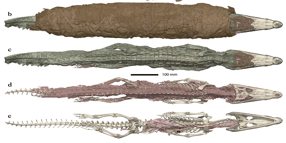
In the project, my job is to use artificial intelligence to automatically recognise and colour in the mummy parts. This year-long project will bring down the time spent segmenting objects from a few months to a few days or hours. It will allow the archaeologists to study populations of animal mummies rather than individuals, which will give us a better understanding of this strange but fascinating practice.
The ASEMI project is a collaboration between the University of Malta and the European Synchrotron Radiation Facility, and has received funding from the ATTRACT project funded by the EC under Grant Agreement 777222. The project is led by Prof. Johann A. Briffa (Department of Communications & Computer Engineering, Faculty of Information & Communication Technology), and a team of researchers from the Data Science Research Platform, University of Malta, and four researchers at the ESRF.


Comments are closed for this article!