Author: Dr Claude J. Bajada
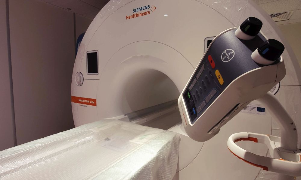
Mater Dei Hospital is a stone’s throw away from the University of Malta. In a new and exciting collaboration between the two institutions, the university invested in a Magnetic Resonance Imaging (MRI) scanner for brain research, tumour detection, and many other crucial studies.

An MRI machine is a very large, very strange digital camera. Instead of detecting light, MRI scanners use powerful electromagnetic fields (some about 45,000 times the strength of the Earth’s field) to give energy to the body’s water. It is that energy that is detected and converted into a digital image.
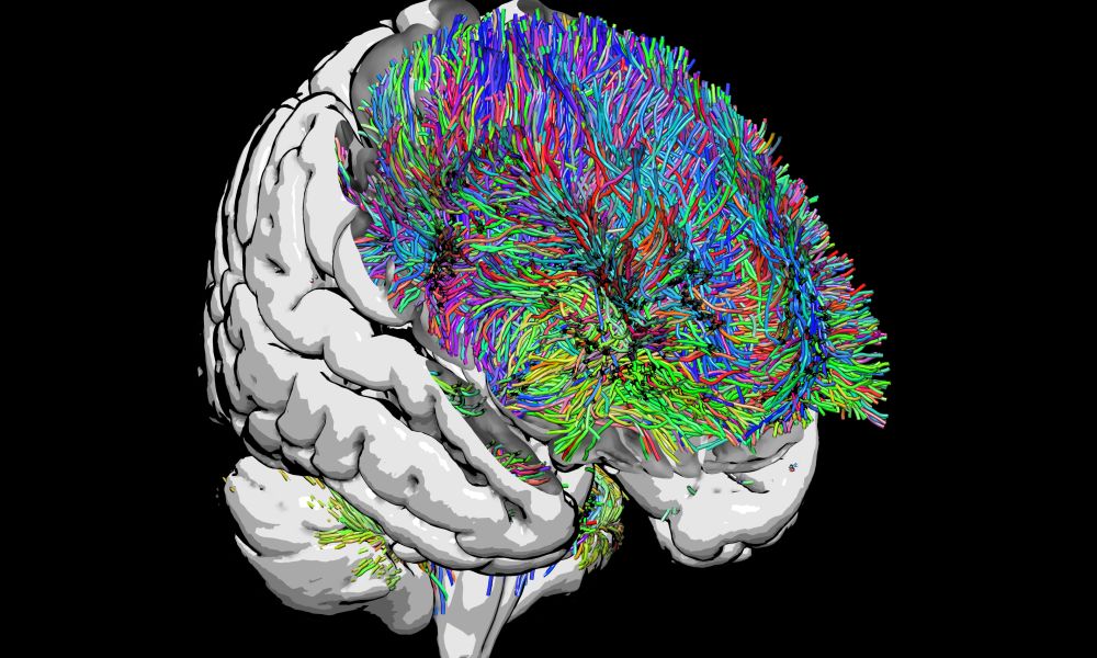
The complex process of obtaining an image is what makes MRI so useful. There are many ways to tweak the input and intermediary steps before obtaining this image, allowing scientists and doctors to obtain very detailed images of a person’s insides and to visualise things that sound like science fiction. An MRI scanner can identify tumours in the liver, lungs, or breast. But it can also be used to reconstruct the connections in the brain or to visualise the fluctuations of neural activity.

UM scientists, as part of a cross-faculty and centre working group, are devising novel approaches to analysing MRI data. They are asking questions that can only be answered by looking deep inside the human body. The joint project between the hospital and the university will not only contribute to the diagnosis of diseases — it will also enable cutting-edge science and ultimately improve Malta’s medical imaging abilities for all.
The device will be housed at the hospital’s Medical Imaging Department. The scanner acquisition is funded through the Transdisciplinary Research and Knowledge Exchange (TRAKE) Complex, co-financed through the European Regional Development Fund 2014 – 2020 (ERDF.01.0124). Claude J. Bajada is a member of the University of Malta MRI Working Group.


Comments are closed for this article!