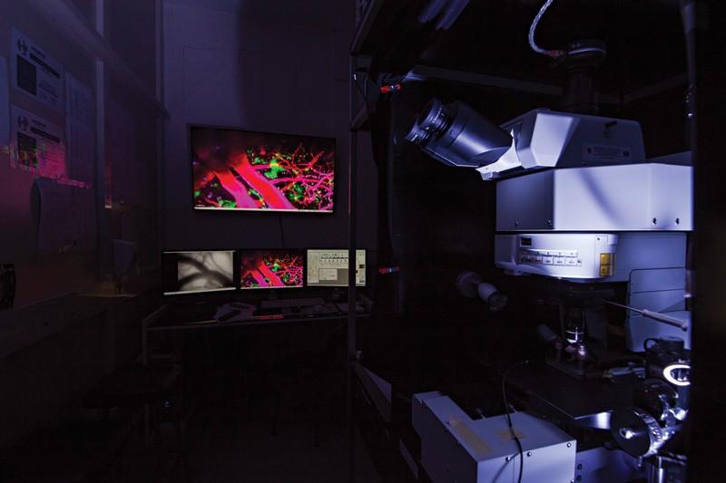
| The Olympus Fluoview FV1000-MPE using ultrashort pulsed IR laser |
|
QUICK SPECS
|
In the last 25 years, two-photon excitation microscopy paved the road for the most significant advance in bio-imaging. This year, four scientists (Winfried Denk, Arthur Konnerth, Karel Svoboda, and David Tank) were awarded the prestigious Grete Lundbeck European Brain Research prize for its invention and development. The method has transformed brain research since it allows real-time examination of the brain’s finest structures. It is powerfully used to investigate stroke, Alzheimer’s disease, migraine, and epilepsy.
The University of Malta’s microscope combines ultrashort-pulsed infrared laser to excite fluorescent molecules up to a depth of 1mm in the rodent brain. The technique allows flexible detection of the brain’s geometries and can look 5–20 times deeper than other types of fluorescent microscopes. The customised setup can perform live imaging to create 3D brain images.
At the University the instrument is used by six scientists on a daily basis with two foreign collaborators in fields which include the evolution of stroke, brain-blood flow dynamics, neurovascular coupling, epilepsy, potassium channel physiology, and white matter injury.
The microscope can aquire images through four simultaneous color channels at 30 frames per second. During imaging of small anaesthesized animals, the microscope is equipped for monitoring vital signs. The instrument is housed in a temperature of 22°C and <30% humidity controlled environment adjacent to a surgical preparation suite and imaging workstation. Two-photon microendoscopy has started to find clinical applications in cancer. There are ongoing developments to image deeper brain structures.


Comments are closed for this article!