

Ingrid Vella writes about her career in medical imaging and how she ended up with pictures of her kidneys.
From a young age, I have shared a passion for both engineering and medicine. I was intrigued by how stuff works: how a TV receives signals, how an aeroplane flies, how a bulb lights up, how humans move their arm. My father, a physics teacher, answered most of my questions but this only fuelled my curiosity to read for an Honours Degree in Electrical Engineering at the University of Malta. During my degree I spent one semester on an Erasmus exchange at the University of Nottingham.
In my final year project in Malta, I analysed ElectroEncephaloGraphy (EEG) data that can detect a person’s brain waves to decipher which limb they are trying to use, which is useful, for example, to paralysed people who need to move a robotic arm. This study encouraged me to read for a Masters in Biomedical Engineering at Imperial College, London in 2011.
During my M.Sc., I worked on a research project in the Brain and Behaviour Lab at Imperial. The aim was to devise an objective and quantitative way to determine if a person had problems with movement. To this end, I recorded 300 hours of movement data using a motion capture suit, and analysed the similarities and differences in movement among healthy people against patients suffering from Parkinson’s Disease.
Following my Masters Degree, I started a Doctorate in MRI (Magnetic Resonance Imaging) Physics at the University of Nottingham. MRI is a way to image parts of the body without the need for messy surgery. I chose the University of Nottingham because it is a pioneer university in MRI research. Sir Peter Mansfield, a professor at the University of Nottingham, was awarded the Nobel Prize for Medicine in 2003, shared with Paul Lauterbur, for discoveries in MRI. The University of Nottingham boasts cutting edge technology.
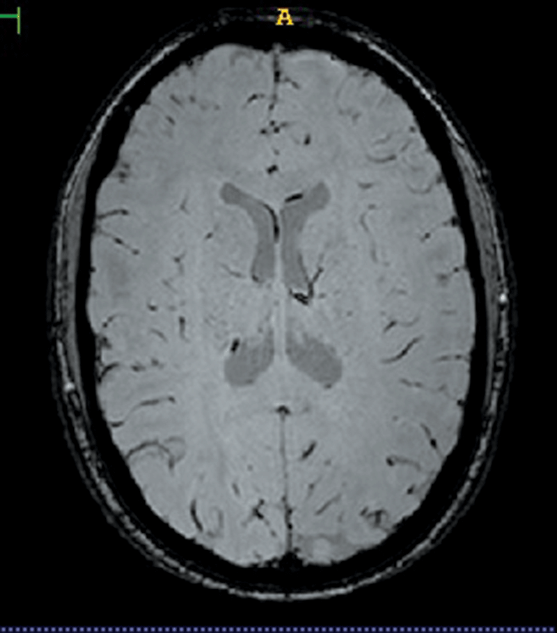
In Nottingham, I am trying to improve MRI hardware that is used to image the human brain at Ultra High Field (UHF). The typical MRI magnets used in hospitals are three Tesla strong, whereas MRI magnets at UHF are at least seven Tesla strong. The higher the magnetic field the better the resolution of the image. When a person’s head is subjected to the magnetic field from an MRI magnet, the magnetic field becomes inhomogeneous, which is not good. The aim of my project is to find ways by which to keep the field as homogeneous as possible during imaging of the human head.
I am also working on a project using MagnetoEncephaloGraphy (MEG) aimed at creating a new way to image using this technology. MEG is a non-invasive technique that is typically used to measure the magnetic fields generated by the neuronal activity of the brain.
Since my colleagues and I need to test our research, it is inevitable that we volunteer as each other’s subjects. I enjoy taking part in my colleagues’ experiments, and I find that by participating in them, I learn more about their research projects. Thanks to these experiments, I have images of my brain, spine, and kidney, as well as a cool video of my heart pumping. I predict that by the end of my Ph.D., I would have gathered multiple images of all my organs! These are some strange benefits of embarking on a research career in medical imaging.


Comments are closed for this article!