
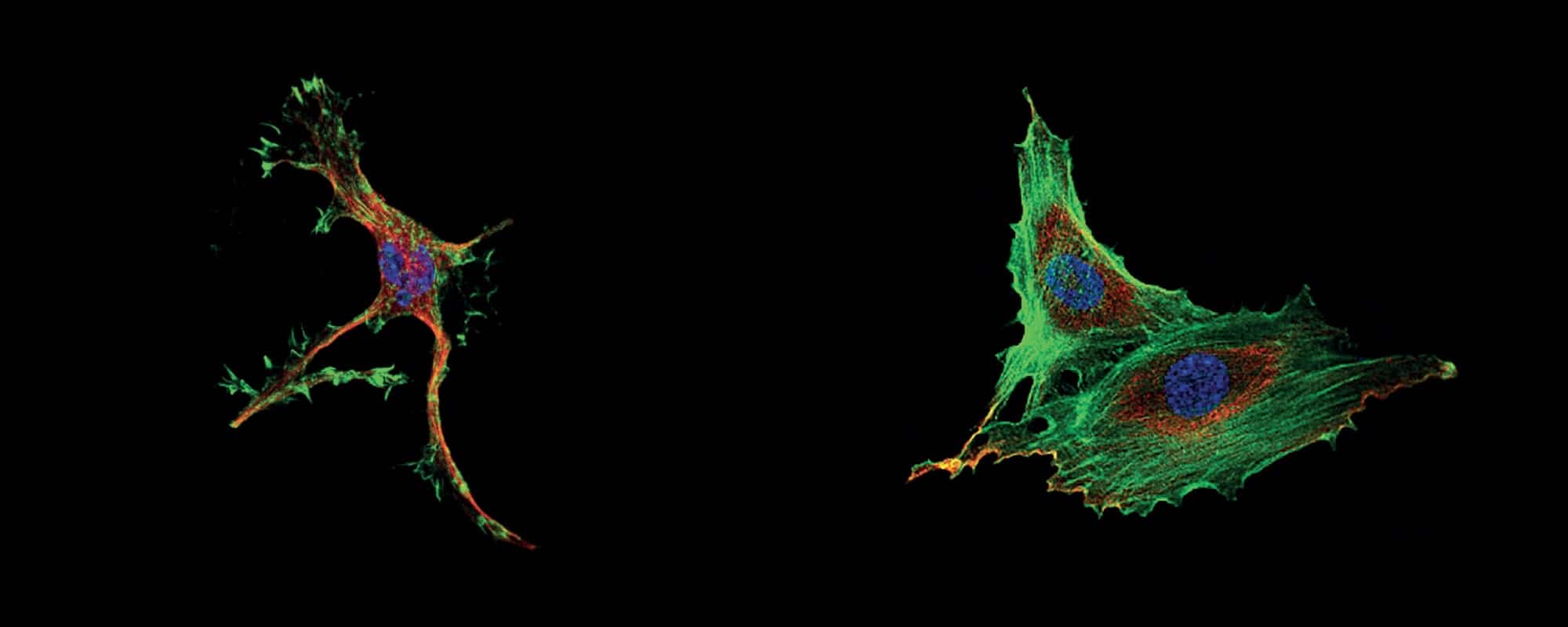

Imagine swallowing a substance which suddenly causes your entire skeleton to collapse in on itself: all your bones squashed and squeezed together in the most unnatural positions. What a tragedy that would be! Luckily, such a toxin does not exist for humans, but it does at the cellular level. In fact, I (Tymoteusz Piasecki), together with my colleagues, supervised by Prof. Zenon Rajfur and his Ph.D. students at the Jagiellonian University of Kraków in Poland, focused on the effects of such a toxin (Cytochalasin D) on cells.
The skeletons of cells (the cytoskeleton) are made up of roughly three parts: threads called intermediate filaments, which help keep the shape of the cell; tubes called microtubules, which also help keep the structure of the cell in addition to helping transport materials inside the cell; and another type of thread, actin, which is partly responsible for cell movement. In fact, the toxin we used actually disrupts the actin part of the cytoskeleton.
I first video-recorded our cells without the toxin added. They looked quite healthy, able to divide and roam around the dish. However, upon adding a high concentration of the toxin Cytochalasin D, cell movement suddenly stopped as the cells became smaller and assumed spikier shapes. Next, I killed and stained the cells, not out of cruelty, but to observe them under a more complex confocal microscope in fluorescent light (see pictures). The cells with the toxin showed major abnormalities in the actin part of the cytoskeleton, which differed for different concentrations of the toxin. During the project, I should add that it was necessary to supply the cells with the correct food, making sure they did not overgrow while also keeping everything sterile to prevent bacterial infections.
I then analysed the recordings to obtain data such as changes in direction of movement, velocity, area, etc. and look for any patterns. One such numerical outcome was that, upon adding the toxin, the area taken up by the cells decreased, pointing to large changes in the cells’ internal structures. These results are to be used in future projects by the researchers.
In general, toxins which affect the cytoskeleton (such as Cytochalasin D itself) are, in fact, used to study the cell’s skeleton, but some are also used as pesticides. More importantly however, these toxins are used in certain medicines as well as chemotherapeutic agents, which makes this field of research truly an important one.
The following colleagues worked with me on the project: Daria Chigarina, Agata Cournuaud, Dawid Marcisz, and Piotr Musiał, supervised by the following Ph.D. students: Armin Mirzaki Ebrahimi, Asia Hajduk, and Tomasz Kołodziej.
Tymoteusz Piasecki is currently following a course leading to Doctor of Medicine and Surgery at the Faculty of Medicine and Surgery, University of Malta. The internship at the Jagiellonian University in Kraków, Poland was organised independently by the author.



Comments are closed for this article!